Bacteria are tiny microorganisms that have the most amazing strategies to cause disease and survive in their environments. They need nutrients, the right amount of oxygen, light and temperature to survive but what happens when their environment and conditions needed for their existence change? Do they just die?
My job as a research microbiologist is to focus on the ‘bad bacteria’ – the ones that make people sick. In this article, I am going to tell you about some of the tools and tricks that bacteria have to help them survive in the environment and how these tools help the bacteria to cause disease in humans.
Bacteria come in different shapes and sizes, some are rod shaped (bacillus), some are spherical (coccus), some are comma shaped (vibrio), and some are spiral shaped (spirilla). Bacteria can occur in pairs such as diplococci (pneumococcus), in chains such as streptobacilli or streptococci, grape-like bunches such as staphylococci, packets of eight or more are sarcinae and groups of four in a square shape are tetrads. Bacillus bacteria can also form palisades where they resemble a picket fence.
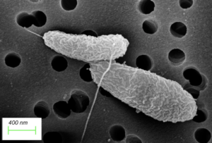
Figure 1: A scanning electron micrograph picture of two Vibrio bacteria together. Vibrio bacteria are rod-shaped and have a single flagellum. The long hair-like structure protruding out of the cell is called the flagellum. Photo source: Dr Sariqa Wagley, University of Exeter.
All about my favourite bacteria…
Before I tell you about some of those tricks and tools, let me introduce you to my favourite bacteria. It is called Vibrio. It loves warm temperatures and salty environments. It lives in the marine environment, in seawater or inside shellfish (molluscs), such as oysters and mussels and in crustacean, such as crabs and prawns during the summer months. Vibrio are rod shaped bacteria and they have stealth-like weapons to help them survive in the environment. When people eat shellfish that are contaminated with Vibrio bacteria, the bacteria can use their weapons to cause diarrhoea and vomiting – this is called gastroenteritis. Vibrio bacteria are the leading cause of seafood-associated gastroenteritis worldwide.
Infecting humans
Here are three tools that Vibrio bacteria have to help them to enter the human gut and cause gastroenteritis.
Flagella – (singular is flagellum, plural is flagella).
Flagella look like strands of hair, and they aid bacteria to swim and attach to surfaces. In the turbulent marine environment, bacteria use the flagella to swim, to attach to surfaces such as the insides of shellfish where they can settle and grow. Unfortunately for us, when Vibrio bacteria are eaten by humans, the bacteria use their flagella to swim and make it to the gut, there they further use the flagella to attach to the lining of the gut.
Toxins
Toxins are chemicals produced by the bacteria that can cause a reaction in your body. Once the Vibrio bacteria use their flagella to attach to the gut lining they then produce a type of toxin called enterotoxins. Vibrio bacteria release these enterotoxins only once they enter the human gut. Some Vibrio toxins enter the blood cells and cause them to burst open releasing blood into the lower intestine causing bloody diarrhoea as a symptom of infection. Some Vibrio toxins are released in the gut causing water and salt imbalances, which leads to a rice-water like diarrhoea being produced; this diarrhoea is called cholera and caused by a particular species of Vibrio called Vibrio cholerae.
Siderophores
Bacteria need essential nutrients to grow and survive. Vibrio bacteria need iron to grow and they have developed special tools to scavenge iron from their surroundings. One of those tools are called siderophores. Siderophores allow bacteria to take iron from other bacteria, environmental sources or from iron sources in our bodies such as lactoferrin and transferrin. If they cannot get hold of that iron, the bacteria will not survive very long inside us and the infection is limited.
Some stealthy shape shifting tricks
Vibrio bacteria need salt and warm temperatures to flourish and so we often detect them in summer months when the conditions are perfect for them to grow. Vibrio bacteria are not detected in the marine environment in the winter months and it is assumed they die due to the cold temperatures and the lack of nutrients. The following summer the Vibrio bacteria will reappear to inhabit the environment and cause seasonal infections once more. So the question is – do Vibrio bacteria really die in the winter?
In laboratory experiments, we can show that some cells of Vibrio bacteria enter a state of dormancy or a sleep-like behaviour in order to survive cold temperatures and low nutrient levels. If we take those dormant sleeping Vibrio and put them into warm temperatures and nutrient rich conditions they reawaken and reproduce once more. Is this happening in the environment too? Do Vibrio bacteria survive in the winter by hibernating until the following summer?
As a research scientist, the topic of bacterial dormancy is both fascinating and a complex subject to investigate.
So how do I study them?
In the laboratory, I can put Vibrio bacteria into a sleep like state by placing them under cold temperatures and taking away their food sources. Many of these bacteria die in these harsh conditions but I have found that some Vibrio cells enter a state of dormancy.
How do I know this? I use a special piece of machinery called a flow cytometer to look at all the cells as I put them to sleep.
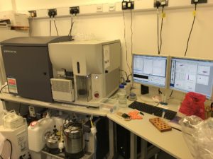
Figure 2: This is a Flow Cytometer that is used to separate cells depending on shape and size. In the left of the picture, the main part of the machinery is on the bench including the flow cell where the light passes through each cell. Underneath the bench, you can see all sorts of tanks containing different chemicals and buffers that are needed to help run the machine. The computer on the right is where the data then appears as the machine analyses the sample. Photo source Dr Sariqa Wagley.
Here is a bit about how it works: I take a sample that has a mixture of cells of varying sizes and shapes (such as a food or water sample) and examine the microscopic contents using the flow cytometer. The machine takes the sample and passes each single particle, one at a time through a small capillary. As each particle passes through the capillary, a light shines upon it and the computer measures the way in which the light scatters as it hits each cell. For example, light will hit and radiate off a round-shaped particle very differently to when light hits a rod shaped particle. The different ways the light scatter means that, I can separate particles and bacteria depending on their shape and size.
Using the flow cytometer, I have found out that dormant Vibrio bacteria look quite different to healthy, alive, awake Vibrio bacteria. When Vibrio bacteria are healthy, they are rod shaped and approximately 1µm in size. When they are lying dormant, they change shape and turn coccoid or spherical in shape. They also balloon out and increase in size. These dormant bacteria utilise nutrients at a reduced rate and are conserving their energy to a bare minimum. They do not divide or grow in this dormant state so they can save energy and resources. This state of dormancy is reversible and if I put the dormant bacteria into warm nutrient rich conditions than they wake up and start growing once more.
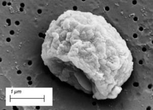
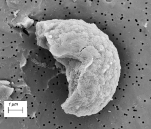
Figure 3: Scanning Electron Microscope pictures of dormant Vibrio bacteria. You can see from Figure 1 what healthy Vibrio bacteria look like and how much they change when they become dormant in this picture. The healthy bacteria in Figure 1 have been put into nutrient deprived media and at cold temperatures to mimic the scenario they may encounter in the environment during the winter. After 40 days in these harsh conditions, they have changed shape to the ones seen in this figure above. The cool thing is that if I place these dormant bacteria into warm nutrient rich environment again they ‘wake up’ and turn back to their healthy form seen in Figure 1. Photo source: Dr Sariqa Wagley, University of Exeter.
Why do I study them?
When microbiologists examine food and clinical samples for the presence of microbes they typically measure the bacteria that are actively growing and dividing. Because dormant cells do not grow and use little energy, they are much harder to detect and evade our conventional microbe detection tests. Researching bacterial dormancy is going to be important in developing new ways to detect these dormant cells so we can detect them before they wake in food and medical samples. If I can detect Vibrio bacteria before they wake up in the summer this could help prevent gastroenteritis infections from occurring in the first place.
I am also investigating if Vibrio bacteria are present naturally in in the environment during the winter. In my future research, I hope to look at these dormant bacteria in nearshore ecosystems inside oysters. I want to find out how they adapt from favourable conditions in the summer to more unfavourable conditions in the winter? What mechanisms do dormant cells use to cope with the changing environment? What tools do they have that allow them to become dormant cells? These are all important questions that I hope to answer one day.
Dr Sariqa Wagley
Research Microbiologist, Exeter University

Download PDF
If you wish to save, or print, this article please use this pdf version »
Learning resource
We have created learning notes to assist students and educators to further investigate the topics covered in this article. You can download the learning resource here »
